What is a cerebral aneurysm?
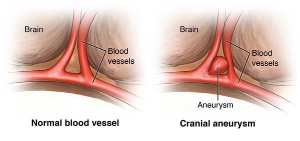
A cerebral aneurysm is a bulge in a weak area of the wall of a brain artery. It puffs out like a small balloon and fills with blood. It is also called an intracranial aneurysm or brain aneurysm. This bulge makes the artery more likely to tear (rupture) in that spot. A cerebral aneurysm occurs more often in arteries at the base of the brain. But it may occur in arteries anywhere in the brain.
What causes a cerebral aneurysm?
Researchers don’t fully know what causes cerebral aneurysms. They are linked to several things. These include:
- Advanced age
- Smoking
- High blood pressure
- Binge alcohol drinking
- Being a woman
- Family history of aneurysms
- Polycystic kidney disease
The main cause of a brain aneurysm is a weakening in the wall of an artery. The pressure of the blood being pumped through the artery can then cause the bulge in that weak area. In certain areas, an aneurysm may have more pressure on it. For example, a place where the artery divides into smaller branches may have more pressure.
How is a cerebral aneurysm treated?
Many factors are considered when making treatment choices. These include:
- The size and location of the aneurysm
- If you have symptoms
- Your age and overall health
- If you have risk factors for aneurysm rupture
In some cases, the aneurysm may not be treated. You may instead be closely watched by a healthcare provider over time. In other cases, surgery may be needed.
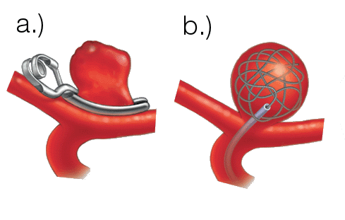
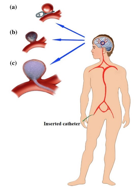
There are 3 main types of surgery for a cerebral aneurysm (Bodaghi et al.) . They are:
· a) Open craniotomy (surgical clipping)
This procedure is done by removing part of the skull to reach the aneurysm. The surgeon places a metal clip across the neck of the aneurysm. This is done to prevent blood flow into the aneurysm bulge. Once the clipping is done, the skull is closed back together.
· b) Endovascular coiling
This is also called coil embolization. It is a minimally invasive method. It is the most common method used to treat cerebral aneurysms. An incision in the skull is not needed. Instead, a catheter is pushed up from a blood vessel in the groin into the blood vessels in the brain. Fluoroscopy (live X-ray) is used to help move the catheter into the head and into the aneurysm. Once the catheter is in place, tiny platinum coils are put through the catheter into the aneurysm. These soft coils are visible on an X-ray. They conform to the shape of the aneurysm. The coiled aneurysm becomes clotted off (embolization). That removes blood flow to the aneurysm to prevent rupture. This procedure is done with general anesthesia, sedation, or local anesthesia.
· c) Foaming
Related topic:

0 Comments